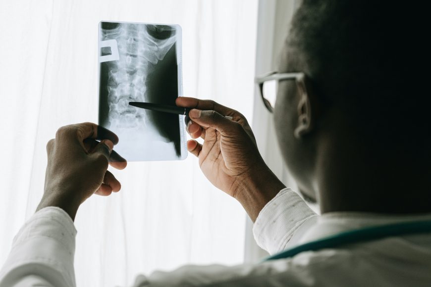
Radiology is the medical discipline that uses medical imaging to diagnose and treat diseases within the bodies of animals, including humans.
Radiographs (originally called roentgenographs, named after the discoverer of X-rays, Wilhelm Conrad Röntgen) are produced by transmitting X-rays through a patient. The X-rays are projected through the body onto a detector; an image is formed based on which rays pass through (and are detected) versus those that are absorbed or scattered in the patient (and thus are not detected). Röntgen discovered X-rays on November 8, 1895 and received the first Nobel Prize in Physics for their discovery in 1901.
Οur Process
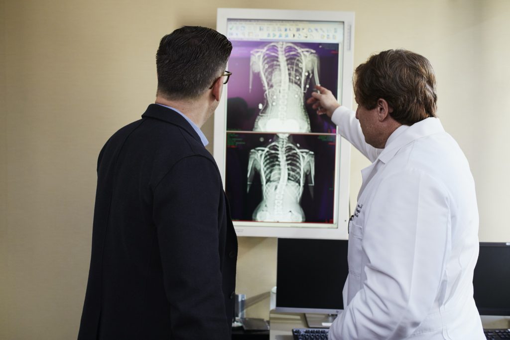
In film-screen radiography, an X-ray tube generates a beam of X-rays, which is aimed at the patient. The X-rays that pass through the patient are filtered through a device called a grid or X-ray filter, to reduce scatter, and strike an undeveloped film, which is held tightly to a screen of light-emitting phosphors in a light-tight cassette. The film is then developed chemically and an image appears on the film. Film-screen radiography is being replaced by phosphor plate radiography but more recently by digital radiography (DR) and the EOS imaging.
In the two latest systems, the X-rays strike sensors that converts the signals generated into digital information, which is transmitted and converted into an image displayed on a computer screen. In digital radiography the sensors shape a plate, but in the EOS system, which is a slot-scanning system, a linear sensor vertically scans the patient.
- Design, implementation, application and evaluation of financial instruments.
- Fund management for the implementation of financial instruments.
- Consulting services for the design, support, funding and implementation of Concession projects or PPPs.
- Independent Expert Review of Investment Plans.
- Design and monitoring of Business Plans and Feasibility Studies.
Plain radiography was the only imaging modality available during the first 50 years of radiology. Due to its availability, speed, and lower costs compared to other modalities, radiography is often the first-line test of choice in radiologic diagnosis. Also despite the large amount of data in CT scans, MR scans and other digital-based imaging, there are many disease entities in which the classic diagnosis is obtained by plain radiographs. Examples include various types of arthritis and pneumonia, bone tumors (especially benign bone tumors), fractures, congenital skeletal anomalies, and certain kidney stones.
Mammography and DXA are two applications of low energy projectional radiography, used for the evaluation for breast cancer and osteoporosis, respectively.
Pricing
From only $20
Based on how quickly you would like your scan, and the location.
Locations
Locations across the country
Based on how quickly you would like your scan, and the location.
Appointments
Same day appointments
Based on how quickly you would like your scan, and the location.
Appointments
Referral by doctor
Based on how quickly you would like your scan, and the location.
Consultants