A variety of techniques are used, depending on the suspected abnormality.
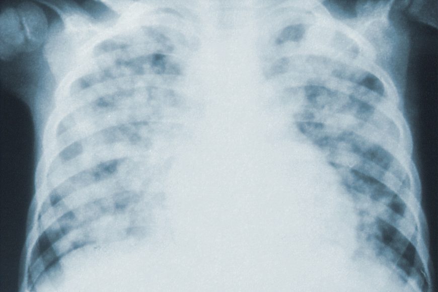
For evaluation of chronic interstitial processes such as emphysema, and fibrosis, thin sections with high spatial frequency reconstructions are used; often scans are performed both on inspiration and expiration. This special technique is called high resolution CT that produces a sampling of the lung, and not continuous images. Bronchial wall thickening can be seen on lung CTs and generally (but not always) implies inflammation of the bronchi
Οur Process
An incidentally found nodule in the absence of symptoms (sometimes referred to as an incidentaloma) may raise concerns that it might represent a tumor, either benign or malignant. Perhaps persuaded by fear, patients and doctors sometimes agree to an intensive schedule of CT scans, sometimes up to every three months and beyond the recommended guidelines, in an attempt to do surveillance on the nodules.
- Design, implementation, application and evaluation of financial instruments.
- Fund management for the implementation of financial instruments.
- Consulting services for the design, support, funding and implementation of Concession projects or PPPs.
- Independent Expert Review of Investment Plans.
- Design and monitoring of Business Plans and Feasibility Studies.
Established guidelines advise that patients without a prior history of cancer and whose solid nodules have not grown over a two-year period are unlikely to have any malignant cancer.
For this reason, and because no research provides supporting evidence that intensive surveillance gives better outcomes, and because of risks associated with having CT scans, patients should not receive CT screening in excess of those recommended by established guidelines.
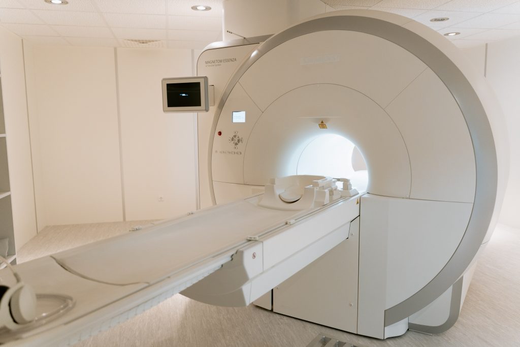
CT scanners use a rotating x-ray tube and a row of detectors placed in the gantry to measure X-ray attenuations by different tissues inside the body. The multiple X-ray measurements taken from different angles are then processed on a computer using reconstruction algorithms to produce tomographic (cross-sectional) images (virtual “slices”) of a body. The use of ionizing radiations sometimes restricts its use owing to its adverse effects. However, CT can be used in patients with metallic implants or pacemakers where MRI is a contraindication.
Pricing
From only $199
Based on how quickly you would like your scan, and the location.
Locations
Locations across the country
Based on how quickly you would like your scan, and the location.
Appointments
Same day appointments
Based on how quickly you would like your scan, and the location.
Appointments
Referral by doctor
Based on how quickly you would like your scan, and the location.
Consultants